ACUTE CHOLECYSTITIS- AI Simulated Case Scenario discussions- Acute Abdomen
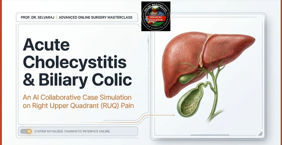
Acute Cholecystitis and Biliary Colic Study Guide
Introduction and Etiology
Biliary colic is caused by a transient obstruction of the cystic duct by a gallstone, which resolves as the stone dislodges .
Acute cholecystitis represents a persistent obstruction of the cystic duct leading to gallbladder inflammation, edema, and potential secondary bacterial infection .
Calculous cholecystitis accounts for 90 percent of cases, while the remaining 10 percent are acalculous .
Risk factors for calculus cholecystitis are often summarized by the 4 Fs: Female, Fat (obese), Fertile (multigravida), and Forty (elderly) .
Acalculous cholecystitis typically affects critically ill patients, those with severe sepsis, or individuals on long-term total parenteral nutrition .
Gallstone formation is linked to the Admirand Triangle, where an imbalance in the ratio of cholesterol, lecithin, and bile salts leads to precipitation .
Clinical Presentation and Comparison
Biliary colic presents as severe, continuous right upper quadrant or epigastric pain that typically begins 1 to 2 hours after a fatty meal .
The pain in biliary colic is episodic and self-limiting, usually resolving spontaneously within 1 to 4 hours .
Acute cholecystitis involves persistent pain lasting more than 6 hours, often accompanied by fever and vomiting .
Murphy’s sign is the most specific clinical finding for acute cholecystitis and is elicited when a patient experiences an abrupt inspiratory arrest during deep palpation of the right hypochondrium .
Patients with acute cholecystitis may also exhibit localized guarding or a palpable tender mass in the right upper quadrant .
Diagnostic Investigations
Right upper quadrant ultrasound is the gold standard imaging modality for both conditions .
Ultrasound findings for acute cholecystitis include gallstones with posterior acoustic shadowing, gallbladder wall thickening greater than 4mm, and pericholecystic fluid .
In biliary colic, the ultrasound confirms the presence of gallstones but the gallbladder wall remains normal .
A HIDA scan is used for equivocal cases; the non-visualization of the gallbladder is diagnostic for acute cholecystitis .
Laboratory findings in acute cholecystitis typically show leukocytosis and elevated C-reactive protein levels .
Abdominal X-rays may reveal radio-opaque stones in only 10 percent of cases or show gas in the gallbladder wall in emphysematous cholecystitis .
Severity Grading (Tokyo Consensus Guidelines)
Grade I (Mild): Inflammation is limited to the gallbladder in a healthy patient with no organ dysfunction .
Grade II (Moderate): Associated with an elevated white cell count above 18,000, a palpable tender mass, or symptoms lasting longer than 72 hours .
Grade III (Severe): Characterized by organ dysfunction involving the cardiovascular, neurological, respiratory, renal, hepatic, or hematological systems .
Management and Treatment
The definitive treatment for symptomatic gallstones is a cholecystectomy .
For biliary colic, the procedure is typically performed as an elective laparoscopic cholecystectomy .
For acute cholecystitis, the standard of care is early laparoscopic cholecystectomy performed within the 72-hour golden window of symptom onset .
Initial emergency management includes keeping the patient nil per os, administering intravenous fluids, and providing broad-spectrum antibiotics covering Enterobacteriaceae and anaerobes .
NSAIDs are preferred for analgesia as they inhibit prostaglandin synthesis and reduce gallbladder mucin production .
Morphine and Pethidine should generally be avoided because they can cause spasms of the Sphincter of Oddi .
High-risk or Grade III patients who are unstable for surgery may require a percutaneous cholecystostomy as a bridge to interval surgery .
Complications
The most feared complications of acute cholecystitis include perforation and biliary peritonitis .
Other serious outcomes include empyema (gallbladder filled with pus), gangrenous cholecystitis, and abscess formation .
Chronic complications may include cholecystoenteric fistulas which can lead to gallstone ileus .
Mirizzi syndrome occurs when a stone in the cystic duct causes extrinsic compression of the common hepatic duct .
Clinical Pearls and Pitfalls
Up to 15 percent of patients with biliary colic may have a normal ultrasound, making clinical diagnosis paramount .
The absence of fever or leukocytosis does not rule out acute cholecystitis, especially in elderly or diabetic patients where the inflammatory response may be blunted .
A gangrenous gallbladder may present with a negative Murphy’s sign because the gallbladder wall has become necrotic .
Delaying surgery beyond the 72-hour window is associated with higher morbidity and increased rates of conversion to open surgery .
Kindly read the below word file on AI collaborative simulated case scenario discussions on Acute Appendicitis, Take home messages, 5 clinical pearls, 5 clinical pitfalls and 10 MCQS on Acute Cholecystitis.
Comments